Nir Research Group
DNA Nanomachine Lab
Ben Gurion University of the Negev
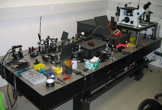
Free Diffusion-smFRET-ALEX
In this setup we are able to measure picomolar concentrations of fluorescently labeled samples which freely-diffuse in and out of the confocal spot. Briefly, an alternating donor-excitation laser (Dex) and an acceptor-excitation laser (AlEX) are focused via the objective to create a diffraction limited spot (A). The donor dye absorbs a photon that originated from the Dex laser and either emits a photon or transfers the energy to the acceptor dye, which, in turn, emits a photon. Alternatively, the acceptor dye directly absorbs a photon that originated from the AlEX laser and then emits a photon. The emitted photons are collected by the objective, split based on their wavelengths, and detected by a single photon detectors (APD). (B) Separate bursts of photons, each corresponding to a single molecule event, are detected. (C) Schematic representation of E and S histogram. (C1) For each burst, E (energy transfer Efficiency) and S (Stoichiometry) are calculated and the results are placed in an E/S 2D histogram. (C2) S-projection reports on the complex structural integrity. (C3) E-projection, in this example, of population located between S=0.4 and S=0.3, (corresponding to one donor and two acceptors attached to a single molecule).

Microfluidics-based single-molecule FRET
TIRF setup: The excitation lasers are focused on the side of the back focal plane of a high numerical aperture objective, creating an evanescent field of ~100 nm, thereby reducing the background fluorescence. A low concentration of fluorescently labeled molecules is immobilized on a coverslip surface via biotin-avidin chemistry, and a flow cell allows exchanging buffer or adding of external molecules.
An integrated microfluidic device is mounted on a single-molecule total internal reflection fluorescence (sm-TIRF) setup. The device includes 16 input channels, which convey solutions containing the molecular components for motor immobilization, assembly, and operation. The pneumatic valves that regulate flow are embedded in the device in a design that enables the rapid and precise computer-controlled exchange of small-volume solutions at a programmable and reproducible sequence and timing.


Defocusing Method for Angle-Resolved Imaging of Gold Nanorods
Defocusing setup: (A) the laser light is circularly polarized by a wave plate (WP), reflected by a silver “dot mirror” (DM), and focused on the center of the back focal plane of a high numerical aperture objective. A low concentration of AuNR-connected molecules is immobilized on the surface with biotin-avidin chemistry and a flow cell allows exchanging buffer or adding external molecules. Light is scattered by the AuNR as a PSF of, among other parameters, its 3D angle which is detected by collecting 1 µm-defocused light with a camera. (B) schematic of the rotation of our DNA-origami “Disk” rotor with an AuNR on top. (C) Rotation angles are extracted from analysis of the recorded movies.

